Exicor® MicroImager™
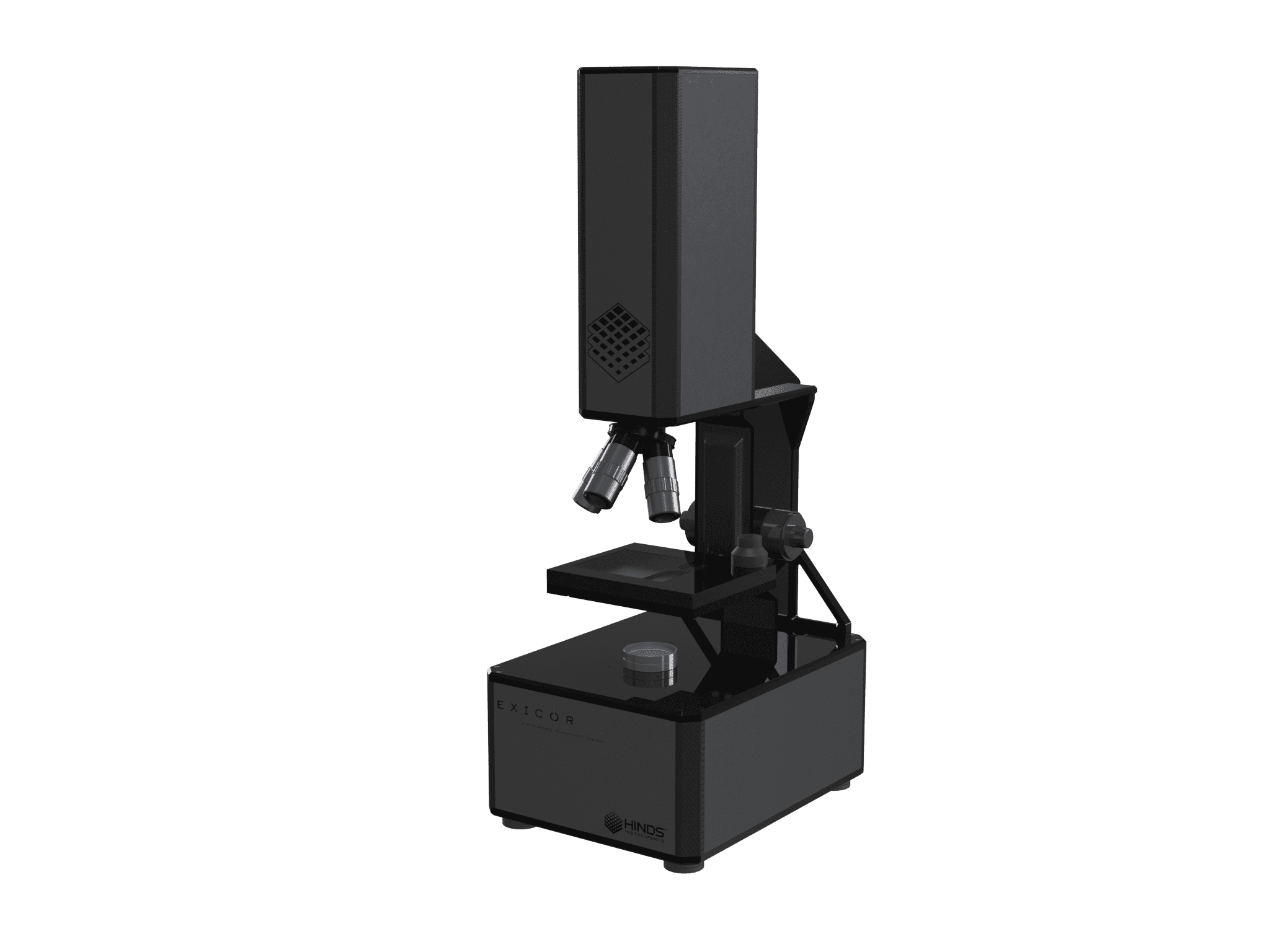
Exicor® MicroImager™
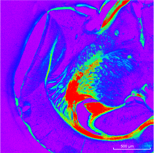
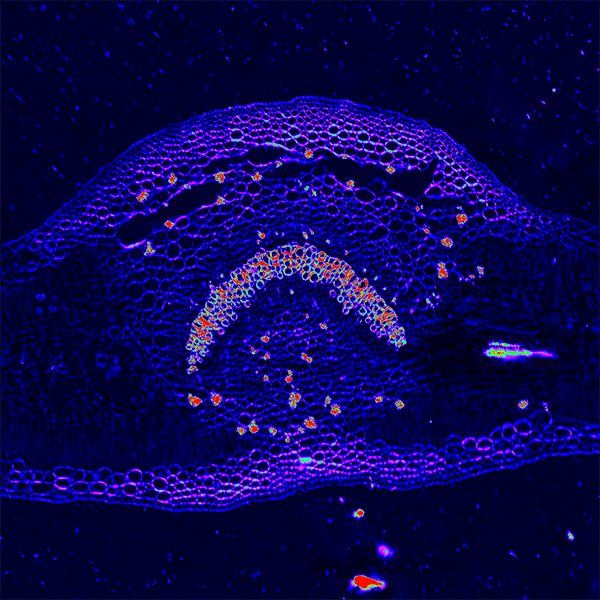
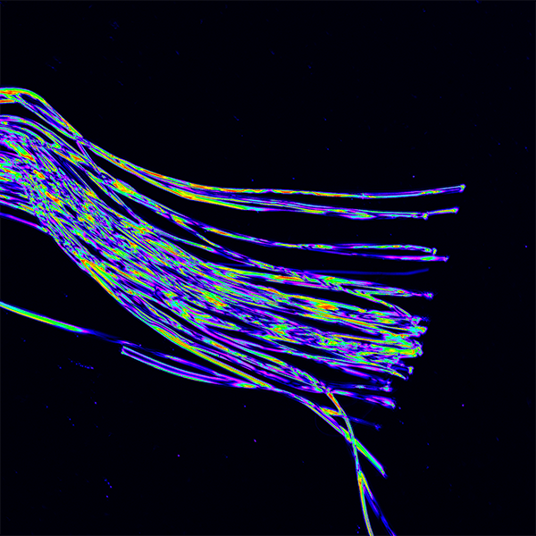
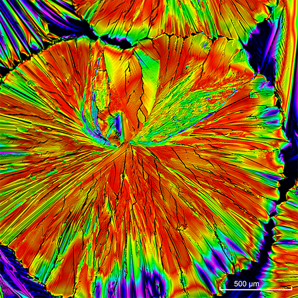
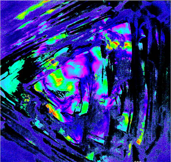
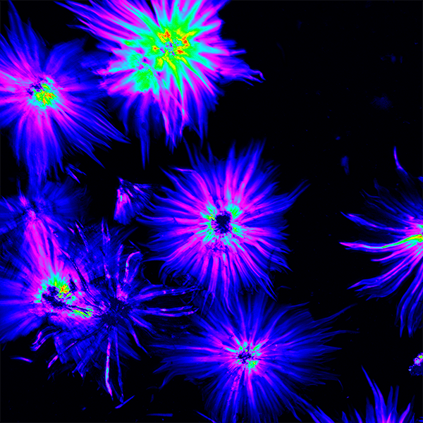
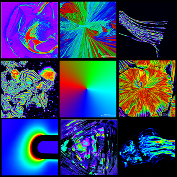
The Exicor MicroImager offers researchers in academia and industry the ability to evaluate birefringence of both biological and industrial materials.
Request QuoteThe Hinds Birefringence Imaging Microscope is ideal for measuring birefringence in biological structures, glass, crystals, and many other organic and inorganic samples.
See below for features and specifications.
System Details
Custom configurations and accessories are available for these models. Please contact a representative for more details.
Features
- No dyes or fluorescent labels required
- Measures retardation, angle, and intensity
- Images retardation, angle, and intensity
- Allows color maps to be customized by user for optimal display of data
- Allows maximum, average, and standard deviation over the entire image and in user-selected areas or lines
- View data by intensity, birefringence/retardation, angle, or a combination of these
- Histograms for statistical analysis
- User calibration
- Export of data in .csv and binary formats
- Export of data from user selected areas
- Export images in png format
- Compatible with third party analysis tools
- Phase unwrapping technology is included with every MicroImager.
- For highly birefringent samples, the Exicor® MicroImager™ has the ability to measure up to 3500nm with 4 color configuration and up to 2400nm with 3 color configuration.
Specifications
| Retardation Repeatability1 | ≤ 0.5 nm (3 sigma) | |
| Light Source Wavelengths | Red | |
| Orange | ||
| Green | ||
| Blue | ||
| Retardation Measurement Range | 2 nm - 0.5λ (1 Color) | |
| Phase Unwrapping to 3500 nm | ||
| Measurement Speed2 | Nominal 15 sec | |
| Dimensions: Footprint | 36.1 cm x 26.5 cm | |
| Height | 77.1 cm | |
| Stage Travel (x, y) | 75 mm, 56 mm | |
| Resolution and Field of View | ||
| Imager | Pixel Resolution | 2464 px x 2056 px |
| Bit-Depth | 12 bits | |
| Objective3 | Resolution, µm | Field of View, mm |
| 2X | 2 | 4.941 x 4.123 |
| 5X | 0.74 | 1.83 x 1.53 |
| 10X | 0.36 | 0.876 x 0.731 |
| 20X | 0.18 | 0.437 x 0.364 |
- 1 Magnitude above 2nm
- 2 2X, 5X, 10X , 20X Noise Floor ~2nm
- 3 1 color with 4 averages
Resources
Videos
Exicor® MicroImager™
Product Manuals
Product Bulletin
Dimensions
Applications
Worldwide Offices
Africa
Hinds Instruments, Inc.
7245 NE Evergreen Pkwy
Hillsboro, OR 97124
USA
Phone: 1.503.690.2000
Fax: 1.503.690.3000
Email: sales@hindsinstruments.com
Americas
Hinds Instruments, Inc.
7245 NE Evergreen Pkwy
Hillsboro, OR 97124
USA
Phone: 1.503.690.2000
Fax: 1.503.690.3000
Email: sales@hindsinstruments.com
India
SciTech Incorporations
D-81 Patel Nagar 2nd
Ghaziabad
Uttar Pradesh 201001
INDIA
Email: sales@scitechincorp.com
URL: www.scitechincorp.com
Japan
Tokyo Instruments Inc.
Attn: Mr. Toshihiko Masuda
T.I.Building,
6-18-14 Nishikasai Edogawa-Ku,
Tokyo 134-0088
JAPAN
Phone: +81 3 (3686) 4711
Fax: +81 3 (3686) 0831
Email: sales@tokyoinst.co.jp
Website: https://www.tokyoinst.co.jp
PR China
OPCrown Photonics Co., Ltd
Room 1006, No.7, Shilong Innovation Building
Yard 98, Lianshihu West Road
Mentougou District, Beijing
Phone: 010-68214292
Fax: 010-68214191
Email: sales@opcrown.com
Sales Director: Brenda Ran
Phone:+86 13911655913
Email: mengqing.ran@opcrown.com
URL: www.opcrown.com
Bei Jing Office
Su Zhou Office
Wu Han Office
Cheng Du Office
Xi An Office
Shen Zhen Office
Bright Stars Technologies Co., Ltd – China HQ
Room 427, Building 12,
No. 55 Shuiyun Road,
Nanhui Xincheng Town, Pudong New Area 201306 Shanghai
Phone: +86-21-51083793
Fax: +86-21-34241962
URL: www.auniontech.com
South Korea
Opto Mechanical Associates (OMA) Co.
405-11,
Goong-dong,
Yusung-gu Daejeon 305 335
KOREA
Phone: +82 42 822 9501-3
Fax: +82 42 822 9504
Email: mail@omacom.co.kr
Website: https://www.omacom.co.kr
Taiwan
Titan Electro-Optics Co., Ltd.
14F., No. 19-11, Sanchong Rd.
Nangang Dist., Taipei City 115, Taiwan
Phone: +886-2-2655-2200 ext. 186
Fax: +886-2-2655-2233
Email: sales@teo.com.tw
URL: www.teo.com.tw
Australia
Hinds Instruments, Inc.
7245 NE Evergreen Pkwy
Hillsboro, OR 97124
USA
Phone: 1.503.690.2000
Fax: 1.503.690.3000
Email: sales@hindsinstruments.com
Austria
Acal BFi Germany GmbH
Oppelner Straße 5
82194 Gröbenzell
GERMANY
Contact: Robert Kardinal
Phone: +49 (0) 8142 6520 119
Fax: +49 (0) 8142 6520 190
Email: robert.kardinal@acalbfi.de
Website: http://www.acalbfi.de/photonics
Belarus (Research Components)
SPECTROPOL
ul. Trakt Lubelski 271 G
04-667 Warszawa
POLAND
Phone: +48 22 617 67 17
Fax: +48 22 617 67 97
Email: biuro@spectropol.pl
Website: http://www.spectropol.pl/
Czech Republic (Research Components)
SPECTROPOL
ul. Trakt Lubelski 271 G
04-667 Warszawa
POLAND
Phone: +48 22 617 67 17
Fax: +48 22 617 67 97
Email: biuro@spectropol.pl
Website: https://www.spectropol.pl
Denmark
Acal BFi Nordic AB (Denmark)
Jernbanegade 23 B
DK-4000 Roskilde
DENMARK
Phone: +45 (0) 7026 2225
Fax: +45 (0) 7026 2221
Email: info@acalbfi.dk
Website: https://www.acalbfi.com/se
Estonia (Research Components)
SPECTROPOL
ul. Trakt Lubelski 271 G
04-667 Warszawa
POLAND
Phone: +48 22 617 67 17
Fax: +48 22 617 67 97
Email: biuro@spectropol.pl
Website: https://www.spectropol.pl
Finland
Acal BFi Nordic AB (Finland)
Teknobulevardi 3-5
FI-01530 Vantaa
FINLAND
Phone: +358 (0) 207 969 770
Fax: +358 (0) 207 969 771
Email: info@acalbfi.fi
Website: https://www.acalbfi.com/se
France
Acal BFi France SAS
1 allée de la Chartreuse
91080 Evry-Courcouronnes
FRANCE
Contact: Jocelyn TANAÏS
Phone: +33 (0) 1 60 79 59 06
Fax: +33 (0) 1 60 79 89 01
Email: photonique.fr@acalbfi.fr
Website: https://www.acalbfi.com/fr
Germany
Acal BFi Germany GmbH
Oppelner Straße 5
82194 Gröbenzell
GERMANY
Contact: Robert Kardinal
Phone: +49 (0) 8142 6520 119
Fax: +49 (0) 8142 6520 190
Email: robert.kardinal@acalbfi.de
Website: https://www.acalbfi.com/de
Ireland
Acal BFi UK Limited
Room 1.09
Challenge House
Sherwood Drive
Bletchley
Milton Keynes
MK3 6DP
UNITED KINGDOM
Phone: +44 (0) 1189 788 878
Fax: +44 (0) 1908 221 110
Email: sales-uk@acalbfi.co.uk
URL: www.acalbfi.com/uk
Acal BFi UK Limited
3 The Business Centre
Molly Millars Lane
Wokingham
Berkshire
RG41 2EY
UNITED KINGDOM
Phone: +44 (0) 1189 788 878
Fax: +44 (0) 1189 776 095
Email: sales-uk@acalbfi.co.uk
URL: www.acalbfi.com/uk
Italy
Acal BFi Italy S.r.I.
Via Cascina Venina n.20
20090 Assago
Milan
ITALY
Phone: +39 (02) 53583.1
Fax: +39 (02) 53583201
Email: sales-it@acalbfi.it
URL: www.acalbfi.com
Laser Optronic SRL
Via Bernardo Quaranta, 57
20139 Milano (MI)
ITALY
Phone: +39 (02) 574651
Fax: +39 (02) 57410127
Email: lop@laseroptronic.it
Latvia (Research Components)
SPECTROPOL
ul. Trakt Lubelski 271 G
04-667 Warszawa
POLAND
Phone: +48 22 617 67 17
Fax: +48 22 617 67 97
Email: biuro@spectropol.pl
Website: https://www.spectropol.pl/
Lithuania (Research Components)
SPECTROPOL
ul. Trakt Lubelski 271 G
04-667 Warszawa
POLAND
Phone: +48 22 617 67 17
Fax: +48 22 617 67 97
Email: biuro@spectropol.pl
Website: https://www.spectropol.pl/
Norway
Acal BFi Nordic AB (Norway)
PO Box 74
3529 Ryse
NORWAY
Phone: +47 3216 2060
Fax: +47 3216 2069
Email: info@acalbfi.no
Website: https://www.acalbfi.com/se
Poland (Research Components)
SPECTROPOL
ul. Trakt Lubelski 271 G
04-667 Warszawa
POLAND
Phone: +48 22 617 67 17
Fax: +48 22 617 67 97
Email: biuro@spectropol.pl
Website: https://www.spectropol.pl
Slovakia (Research Components)
SPECTROPOL
ul. Trakt Lubelski 271 G
04-667 Warszawa
POLAND
Phone: +48 22 617 67 17
Fax: +48 22 617 67 97
Email: biuro@spectropol.pl
Website: https://www.spectropol.pl
Sweden
Acal BFi Nordic AB (Sweden)
Falhagsleden 59
PO Box 1335
SE-751 43 Uppsala
SWEDEN
Phone: +46 (0) 1856 5830
Fax: +46 (0) 1869 6666
Email: info@acalbfi.se
Website: https://www.acalbfi.com/se
Switzerland
GMP SA
Avenue des Baumettes 17
CH – 1020 Renes
Phone: +41 21 633 21 21
Fax: +41 21 633 21 29
Email: info@gmp.ch
Website: https://www.gmp.ch
The Netherlands
Alphen aan den Rijn
Acal BFi Netherlands BV
J.P. Thijsseweg 1e
2408 ER Alphen aan den Rijn
THE NETHERLANDS
Phone: +31 (0) 1 7244 6060
Fax: +31 (0) 1 7244 3414
Email: sales-nl@acalbfi.nl
URL: www.acalbfi.com/nl
Eindhoven
Acal BFi Netherlands BV
Luchthavenweg 53
5657 EA Eindhoven
THE NETHERLANDS
Phone: +31 (0) 4 0250 7400
Fax: +31 (0) 4 0250 7409
Email: sales-nl@acalbfi.nl
URL: www.acalbfi.com/nl
Ukraine (Research Components)
SPECTROPOL
ul. Trakt Lubelski 271 G
04-667 Warszawa
POLAND
Phone: +48 22 617 67 17
Fax: +48 22 617 67 97
Email: biuro@spectropol.pl
Website: https://www.spectropol.pl
United Kingdom
Pro-Lite Technology Limited
Innovation Centre, University Way
Cranfield, MK43 0BT, UK
Contact: Ross Tomlin
Phone: +44 (0)1234 436110
Email: ross.tomlin@pro-lite.co.uk
Website: www.pro-lite.co.uk
Milton Keynes
Acal BFi UK Limited
Room 1.09
Challenge House
Sherwood Drive
Bletchley
Milton Keynes
MK3 6DP
UNITED KINGDOM
Phone: +44 (0) 1189 788 878
Fax: +44 (0) 1908 221 110
Email: sales-uk@acalbfi.co.uk
URL: www.acalbfi.com/uk
Wokingham
Acal BFi UK Limited
3 The Business Centre
Molly Millars Lane
Wokingham
Berkshire
RG41 2EY
UNITED KINGDOM
Phone: +44 (0) 1189 788 878
Fax: +44 (0) 1189 776 095
Email: sales-uk@acalbfi.co.uk
URL: www.acalbfi.com/uk
Middle East
Hinds Instruments, Inc.
7245 NE Evergreen Pkwy
Hillsboro, OR 97124
USA
Phone: 1.503.690.2000
Fax: 1.503.690.3000
Email: sales@hindsinstruments.com
Get A Quote for Systems & Components
Working together, we can discuss the suitability of our technology for your application, including a custom configuration of our base systems if needed.
Featured Article
Fundamentals, Advances, and Artifacts in Circularly Polarized Luminescence (CPL) Spectroscopy
A practical guide to CPL spectroscopy, key to future promising applications.
Upcoming Event
SPIE Photonics West 2025
The leading global conference and marketplace for lasers, biomedical optics, quantum, biophotonics technologies, optoelectronics, microfabrication, displays, and more.

Hinds Instruments © 2026. All Rights Reserved. Built with ♥ by Webfor